Pulmonary Embolism

18 Views

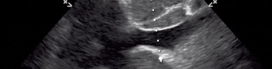
A non-conventional cardiac window, but clearly showing a pleural effusion, the Jellyfish sign of a collapsed lung, and the left ventricle with good contractility along with thickening of the posterior mitral valve leaflet

Pioneer
I think labelling would be the best startegy for pocus sir..as some of us are new to pocus