A non-conventional cardiac window, but clearly showing a pleural effusion, the Jellyfish sign of a collapsed lung, and the left ventricle with good contractility along with thickening of the posterior mitral valve leaflet
80 Views

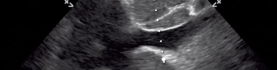
A non-conventional cardiac window, but clearly showing a pleural effusion, the Jellyfish sign of a collapsed lung, and the left ventricle with good contractility along with thickening of the posterior mitral valve leaflet

Diaphragmatic excursion refers to the measurement of diaphragmatic movement during the respiratory cycle using ultrasound. In critically ill patients, it is a bedside, non-invasive method to assess diaphragmatic function and to help predict weaning outcomes from mechanical ventilation.
Technique
Probe placement: A low-frequency (2–5 MHz) curvilinear or phased-array transducer is placed in the subcostal or intercostal region.
Window: The liver (right side) or spleen (left side) serves as an acoustic window.
Mode: M-mode is typically used to quantify movement. The ultrasound cursor is placed perpendicular to the diaphragm dome.

Pioneer
