
Lung ultrasound for the diagnosis of pneumonia in adults: Blue Protocol
- Mazen Kherallah

- Jan 5, 2022
- 2 min read
Updated: Oct 7, 2022
Ultrasound in the intensive care unit is a reasonable gold standard to detect many causes of respiratory failure with a sensitivity and specificity ranging from 90% to 100%. The BLUE-protocol (Bedside Lung Ultrasound in Emergency) was developed at university-affiliated teaching-hospital ICUs in France led by Lichtenstein et al, and is now widely used as a fast and easy tool in the differential diagnosis of acute respiratory failure [1].
The BLUE-protocol combines ultrasound signs (lung sliding, A lines, B lines, and PLAPS) with location, associated with results of a sequential venous analysis (thrombosed versus free veins) to give a clinical diagnosis and etiology of acute dyspnea and respiratory failure.
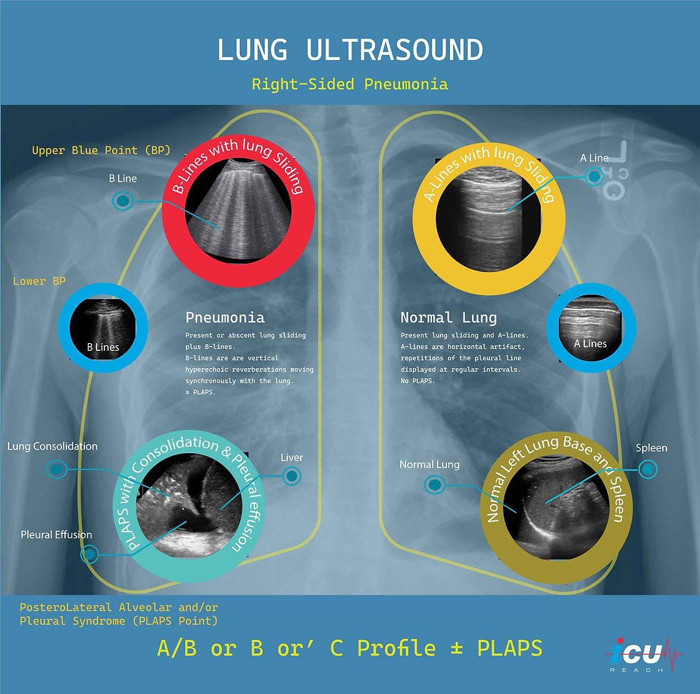
Lung sliding is the shimmering movement of the visceral and parietal pleural surface along with respiration.
A-lines are horizontal artifact result from reverberation of the ultrasound beams off the pleura. They occur at equal intervals and indicate dry interlobular septa (normal lung).
B-lines are discrete laser-like (comets) vertical hyperechoic artifacts extending from the pleura to the edge of the screen and indicate a thickened interlobular septa (pulmonary edema or pneumonia).
PLAPS stands for a PosteroLateral Alveolar and/or Pleural Syndrome representing lung consolidation/collapse and/or pleural effusion.
The combination of these signs results in seven different profiles:
A-profile combines anterior lung-sliding with A-lines bilaterally.
A’-profile when A-lines are present bilaterally with abolished lung-sliding.
B-profile associates anterior lung-sliding with lung-rockets bilaterally.
B’-profile is B-profile with abolished lung-sliding.
C-profile indicates anterior lung consolidation (any size and number) or a thickened, irregular pleural line.
The A/B profile is a half A-profile at one side, a half B-profile at another.
The PLAPS-profile indicates a PosteroLateral Alveolar and/or Pleural Syndrome.
The four profiles that can indicate the presence of pneumonia are A/B-profile, B’-profile, C-profile, or A-profile with clear veins and PLAPS. Ultrasound lacks sensitivity in detecting pneumonia when these profiles are present individually (11-42%) but their absence has an excellent specificity (96-100%). The presence of a combination of these four profiles gives a sensitivity of 89% and specificity of 94% in the diagnosis of pneumonia.
In the above image, CXR shows right sided infiltrate and ultrasound reveals A/B profile (lung sliding with B-lines on the right side and A-lines with lung sliding on the left side) with PLAPS (consolidation with pleural effusion).
References
Lichtenstein DA, Mezière GA. Relevance of lung ultrasound in the diagnosis of acute respiratory failure: the BLUE protocol. Chest. 2008 Jul;134(1):117-25. doi: 10.1378/chest.07-2800. Epub 2008 Apr 10. Erratum in: Chest. 2013 Aug;144(2):721. PMID: 18403664; PMCID: PMC3734893.


Comments