

MRI Images T1, T2, and FLAIR
- Mazen Kherallah

- Jul 31, 2021
- 1 min read
Updated: Jan 7, 2022

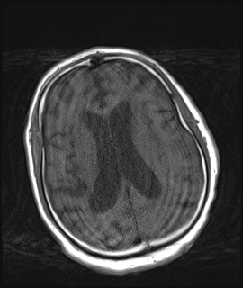
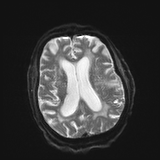

T1-weighted images are produced by using short TE (Time to Echo) and TR (Repetition Time) times. T2-weighted images are produced by using longer TE and TR times.
In general, T1- and T2-weighted images can be easily differentiated by looking the CSF. CSF is dark on T1-weighted images and bright on T2-weighted images.
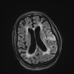
Fluid Attenuated Inversion Recovery (Flair) is similar to a T2-weighted image except that the TE and TR times are very long. By doing so, abnormalities remain bright but normal CSF fluid is attenuated and made dark. This sequence is very sensitive to pathology and makes the differentiation between CSF and an abnormality much easier.
In the attached MRI images, note the confluent hyperintense subcortical and periventricular white matter foci that became very clear on T2/FLAIR as compared to T2 as the CSF became dark contrasting the hypertense foci. These These are no specific and a gadolinium contract is needed to determine etiology.


Comments